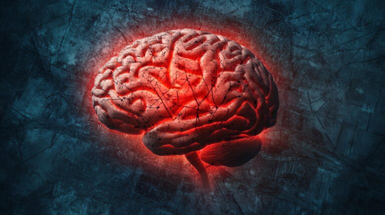
Brain mapping is a powerful and essential technique in modern neuroscience, enabling researchers and clinicians to visualize and understand the intricate details of the structure and function.
By providing a comprehensive view, brain mapping allows us to see how different regions are interconnected and how they work together to control everything from basic bodily functions to complex cognitive processes.
Let us talk about it in greater detail.
The Science Behind Brain Mapping
Brain mapping is a non-invasive method that allows scientists and clinicians to study the brain’s function in detail. It aims to:
- Localize brain functions
- Identify abnormalities
- Develop precise treatment plans
By using various imaging techniques, brain mapping provides a comprehensive view of how different parts interact and contribute to our cognitive and emotional processes.
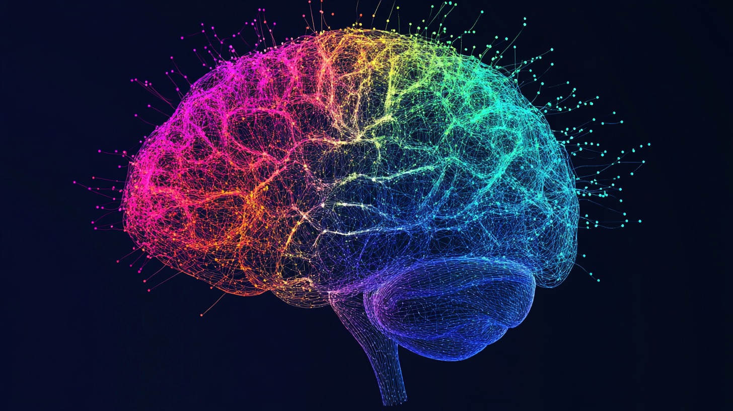
The Human Brain’s Complexity
The human brain is a highly complex organ composed of neurons, lobes, and various types of waves, such as:
- Delta
- Theta
- Alpha
- Beta
Each of these components plays a significant role in function and behavior. Neurons are the building blocks, transmitting signals across different regions.
The brain’s lobes, including the frontal, parietal, occipital, and temporal lobes, are responsible for various functions such as:
- Movement
- Sensation
- Vision
- Memory
Brain waves, which reflect different states of consciousness, are crucial in brain mapping as they provide real-time data on the activity. Understanding these structures and waves is essential for accurately interpreting mapping results.
Brain Mapping Technologies and Methods
Brain mapping is a multidisciplinary field that leverages a variety of imaging techniques to study both the structure and function.
These techniques can be broadly categorized into structural and functional methods, each offering unique insights into how the brain operates and how its various components are interconnected.
Structural Techniques
Structural brain mapping focuses on creating detailed images of the anatomy, allowing for the visualization of its physical structures. Among the most commonly used methods, you will find techniques like:
- CAT (Computerized Axial Tomography) scans
- MRIs (Magnetic Resonance Imaging)
- Diffusion Tensor Imaging (DTI)
CAT Scans provide cross-sectional images using X-rays, which are particularly useful in detecting abnormalities such as:
- Bleeding
- Tumors
- Skull fractures
They are often the first line of imaging in emergency settings due to their speed and ability to reveal acute issues quickly.
MRI Scans offer a more detailed and high-resolution image of the soft tissues without using radiation.
MRI scans can differentiate between various types of tissue, making them invaluable in diagnosing a wide range of neurological conditions.
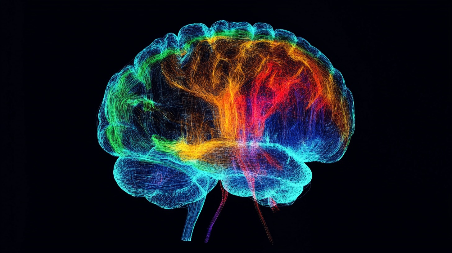
Diffusion Tensor Imaging (DTI) is a specialized form of MRI that maps the diffusion of water molecules.
By visualizing these connections, DTI helps in understanding how various regions communicate and in identifying disruptions in these pathways caused by:
- Trauma
- Stroke
- Neurodegenerative diseases.
These structural techniques are critical in diagnosing and planning the treatment of neurological diseases.
They provide a detailed anatomical roadmap that guides surgeons during operations, ensuring that critical areas of the brain are preserved.
Functional Techniques
While structural techniques focus on the brain’s physical makeup, functional techniques are designed to observe the activity real-time. These methods allow researchers and clinicians to study how different brain regions are involved in various cognitive functions, emotional responses, and behavioral processes.
EEG (Electroencephalography) measures electrical activity in the brain by placing electrodes on the scalp. It is particularly useful for detecting abnormalities in waves, such as those seen in epilepsy or sleep disorders.
EEG provides excellent temporal resolution, allowing for the monitoring of activities on a millisecond-by-millisecond basis, making it ideal for studying the immediate responses to stimuli.
PET (Positron Emission Tomography) scans involve the injection of a radioactive tracer that binds to glucose.
Since active brain regions consume more glucose, PET scans can reveal which areas are involved in specific tasks, such as memory, language, or emotion.
PET is particularly valuable in diagnosing and monitoring neurodegenerative diseases like Alzheimer’s, as it can detect abnormal metabolic activity before structural changes become apparent.
FMRI (Functional Magnetic Resonance Imaging) is a non-invasive technique that measures activity by detecting changes in blood flow.
When a brain region is more active, it consumes more oxygen, leading to an increase in blood flow to that area. fMRI provides both spatial and temporal resolution, allowing researchers to see which parts are involved in specific tasks, such as problem-solving or emotional regulation, while also capturing the timing of these activities.
These functional techniques are invaluable in understanding the dynamic processes. They help in pinpointing the areas responsible for specific functions, which is crucial for developing targeted treatments for neurological and psychiatric disorders.
Brain Mapping Reports and Analysis
Brain mapping results are translated into visual reports that highlight areas of the brain that are over- or under-active. These reports provide a detailed analysis of function, including lobe-specific activity and how it relates to cognitive, emotional, and memory processing.
By interpreting these maps, clinicians can identify patterns of dysfunction that may be contributing to a patient’s symptoms. For instance, hyperactivity in the frontal lobe might be associated with anxiety, while underactivity in the temporal lobe could related to memory issues.
Knowing these patterns is crucial in developing effective treatment protocols that target specific brain regions. The results from brain mapping are vital in guiding the creation of neurofeedback and neuromodulation protocols aimed at normalizing function.
These protocols are designed to address the specific imbalances identified in the maps, helping to restore healthy activity. Supplements and other non-drug interventions may be recommended as part of the treatment plan.
For example, certain nutrients can support health and enhance the effectiveness of neurofeedback. By combining these approaches, clinicians can offer a comprehensive treatment strategy that addresses the root causes of neurological and psychological conditions, promoting long-term recovery.
Effectiveness and Safety of Brain Mapping
Brain mapping has proven effective in clinical practice, with numerous case studies and research findings supporting its use.
It has been shown to improve diagnosis accuracy, guide effective treatment plans, and enhance patient outcomes, particularly in complex neurological conditions.
The safety profile is also well-established, as it is a non-invasive and non-drug procedure. Unlike many medical interventions, brain mapping has no significant side effects, making it a safe option for patients of all ages.
The combination of effectiveness and safety has made brain mapping a valuable tool in modern medicine, particularly in personalized treatment.
The Bottom Line
Brain mapping has revolutionized our understanding of the brain, offering insights that have significantly improved clinical outcomes.
It continues to play a vital role in both research and patient care.